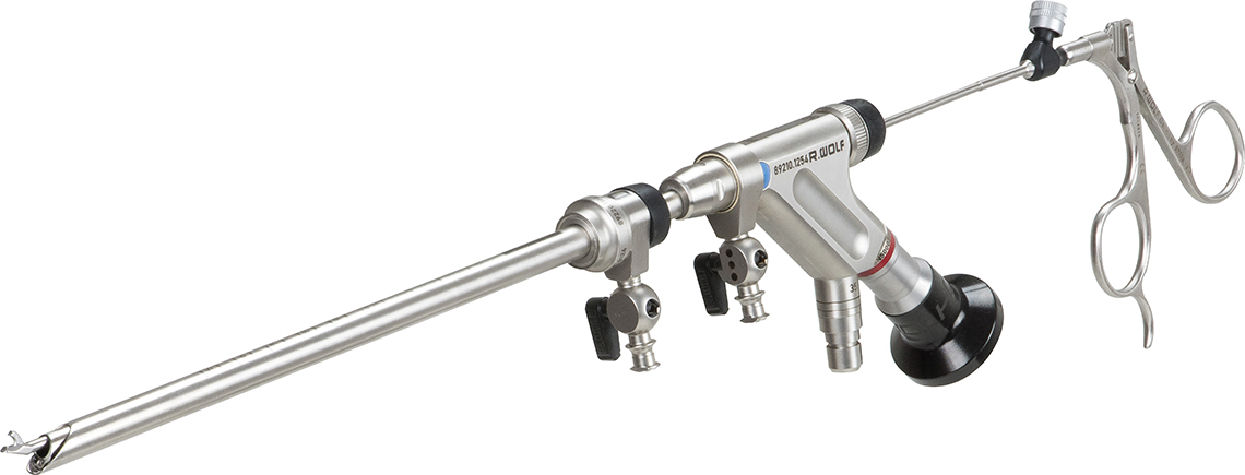
Endoscopic spinal surgery
Back pain is one of the most common reasons for visits to the doctor and hospital. The causes of these complaints can be very diverse, as the human body has a very complex structure and many components, such as muscles, vertebral joints, intervertebral discs, nerves, tumours and fractures can contribute to the development of pain in the back.
Therefore, before any treatment is carried out, the causes must be thoroughly investigated and localised. Once this has been done, various therapeutic and surgical measures are often available.
Exhausting all conservative treatment methods when treating herniated discs and stenoses is of utmost importance. If, despite everything, a surgical intervention is necessary, it is advisable to opt for the most minimally invasive procedure. In the field of lumbar spinal surgery, this is endoscopic spinal surgery.
Full-endoscopic spine surgery offers major clinical and aesthetic benefits such as:
- Patients who underwent endoscopic surgery returned to work after an average of 25 days, while patients in the microscopic group returned after 49 days (p < 0.01).1 Patients often show reduced post-operative pain due to the smaller trauma.
- Significant differences in clinical outcome between endoscopy and microscopy are not observed, but postoperative pain and medication were significantly reduced after endoscopy.1 In addition, the incision is smaller with endoscopy compared with microscopy.
- Complication rates and reoperation rates are the same for both procedures. The recurrence rate of disc herniation is 6.2% and does not differ significantly from the microscopic technique.1.2 Patients who underwent endoscopic surgery have better surgical conditions in the event of a recurrence than those who underwent microscopic surgery due to reduced scarring.
- Operating time: The average operating time in the fully endoscopic technique is significantly shorter than in the microdiscectomy (22 min vs. 43 min, p<0.001). There are no significant differences between the transforaminal and the interlaminar approach.
- Blood loss: In principle, blood loss during full endoscopic surgery is difficult to measure due to constant irrigation. Endoscopic techniques have a lower estimated blood loss than open techniques (3.3 ml vs. 244.9 ml).
- Other advantages of endoscopy are the extended field of view due to 25° optics, rapid rehabilitation of patients and low postoperative costs, reduced anatomical trauma and easier conditions for revision surgery.
1 Ruetten S, Komp M, Merk H, Godolias G. Full-Endoscopic Interlaminar and Transforaminal Lumbar Discectomy Versus Conventional Microsurgical Technique. A Prospective, Randomized, Controlled Study. Spine 2008; 33(9): 931-939
2 Phan K, Xu J, Schultz K. Full-endoscopic vs. micro-endoscopic and open discectomy: a systematic review and meta-analysis of outcomes and complications. Clinical Neurology and Neurosurgery 2017; 154:1-12
PD Dr. Jean-Yves Fournier, Head of Neurosurgery, Valais Hospital, Sion (deutsch)
PD Dr. Christoph Albers, Co-Head of Spine, Inselspital Bern (english)
Prof. Dr. Christoph Siepe, Schön Klinik, München Harlaching, Deutschland
Focus Online on site in the operating room:
Due to the long learning curve for surgeons, endoscopic spinal treatment is still far from being offered in all hospitals. Stöckli Medical has developed a training programme with various experts in this field, which enables surgeons to benefit from a greatly shortened learning curve. This is supported by repeated training on simulation models and the application of various adult-learning principles.
What is the clinical problem?
Herniated discs and spinal canal stenosis occur frequently in patients. Until now, these indications were treated microscopically. With endoscopy, the incision and resulting scar tissue can be reduced to a minimum. The sequester of the herniated discs can be medial and lateral, for which different approaches are used. The patient satisfaction can be increased by the gentle surgical technique.
How does Vertebris work?
The transforaminal and the extraforaminal accesses are made through the intervertebral foramen. With a spinal cannula, the correct position is determined under x-Ray control. By using a dilator, the working cannula can be inserted and placed. The surgery can be done through the endoscope under continuous irrigation.
If the sequester lays more medial, the interlaminar technique is often used. The acces is done through the posterior interlaminar window. The dilator will be placed on the ligamentum flavum and the working cannula is inserted. All access- and working instruments are designed to preserve nerves.


What are the advantages of the Riwo endoscopy system?
The endoscopy system “Vertebris” by RIWO Spine was developed to enable full-endoscopic decompression of the spine. Typical indications for the full-endoscopic surgical technique include herniated discs, spinal cysts and spinal stenosis.
The system offers the following advantages:
- With the adapted lengths of the optics, the most minimally invasive approach can be selected depending on the pathology. Since the system is universal, all accesses can be ensured with minimal set effort
- Wide indication coverage
- 4MHz radio frequency system for increased safety during coagulation
- Fluid management with spine mode to avoid epidural overpressure
- Oval shaft shape of the endoscopes to ensure continuous water outflow
- Specialised instruments for bone resection


